Ecografía de diagnóstico prenatal.
TABLA. Altura Vermis del Cerebelo (mm)+
Los datos base son de Malinger, G., S. Ginath, T. Lerman-Sagie, et al. Prenat Diagn, 2001. 21(8): p. 687-92.


TABLA. Área de los hemisferios cerebelosos (mm)+
Los datos base son de Sherer, D.M., M. Sokolovski, M. Dalloul, et al. Ultrasound Obstet Gynecol, 2007. 29(1): p. 32-7.


TABLA. Diámetro antero-posterior protuberancia (mm)+
Los datos base son de Mirlesse, V., C. Courtiol, M. Althuser, et al. Prenat Diagn, 2010. 30(8): p. 739-45.


TABLA. Diámetro transverso del Cavum Septum pellucidum (mm)+
Los datos base son de Falco, P., S. Gabrielli, A. Visentin, et al. Ultrasound Obstet Gynecol, 2000. 16(6): p. 549-53.


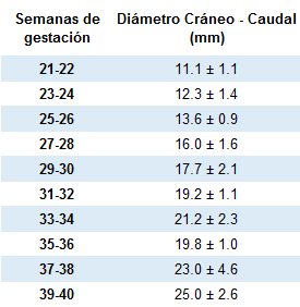
TABLA. Diámetro transverso del cerebelo (mm)+
Los datos base son de Sherer, D.M., M. Sokolovski, M. Dalloul, et al. Ultrasound Obstet Gynecol, 2007. 29(1): p. 32-7.


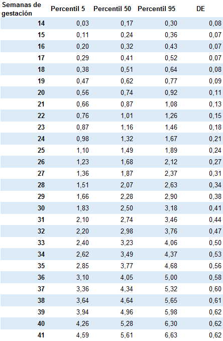
TABLA. Longitud Cuerpo Calloso (mm)+
Los datos base son de Achiron, R. and A. Achiron. Ultrasound Obstet Gynecol, 2001. 18(4): p. 343-7.


TABLA. Riesgo de parto prematuro en función de la longitud cervical+
La presente tabla recoge el riesgo de parto prematuro en función de la longitud cervical a las 20-24 semanas en gestantes asintomáticas con gestación gemelar (según Makrydimas et al., Best Practice Res Clin Obst Gynecol 2014)

TABLA. Longitud cervical efectiva en gestaciones gemelares+
Media y percentiles 5 y 95 de la longitud cervical efectiva en gestaciones gemelares según semana de gestación (medidas en mm).

Datos Crispi et al., Progr Obstet Ginecol; 2004

TABLA. Medidas tercer ventrículo (mm)+
Los datos base son de Sari, A., A. Ahmetoglu, H. Dinc, et al. Acta Radiol, 2005. 46(6): p. 631-5.


TABLA. Perímetro cefálico (mm)+
Los datos base son de Kurmanavicius, J., E.M. Wright, P. Royston, et al. Br J Obstet Gynaecol, 1999. 106(2): p. 126-35.


TABLA. Valores de la translucencia nucal correspondientes a los percentiles 1, 2,5, 5, 10, 50 (mediana), 90, 95, 97,5 y 99 para los distintos valores de CRL+
Los valores base son de Borrell et al. Progr Obstet Ginecol 2006.


Morfometría cardíaca+
Mediciones y cálculo de los diferentes z-score

Schneider C, UOG 2005; 26: 599-605
Información, datos y gráficos - www.interscience.wiley.com/jpages/0960-7692/suppmat/inde.html
(a) Long‐axis view of the left ventricle showing the aortic valve (1) and ascending aorta (2).
(b) Aortic arch view showing the aortic valve (1), ascending aorta (2), descending aorta (3) and inferior vena cava (4).
(c) Short‐axis view, showing the pulmonary valve (1), main (2), right (3) and left (4) pulmonary arteries.
(d) Oblique short‐axis view, showing the pulmonary trunk and the arterial duct (5).
(e) Four‐chamber view, showing the tricuspid valve (1), right ventricular end‐diastolic dimension (2), right ventricular inlet length (3), right ventricular area (dashed line) (4), mitral valve (5), left ventricular end‐diastolic dimension (6), left ventricular inlet length (7) and left ventricular area (dotted line) (8). Ao, aorta; Desc Ao, descending aorta; IVC, inferior vena cava; LA, left atrium; LPA, left pulmonary artery; LV, left ventricle; MPA, main pulmonary artery; RA, right atrium; RPA, right pulmonary artery; RV, right ventricle.
TABLA. Valores doppler normales+


TABLA. Anemia fetal+
Concentración de Hb fetal correspondiente a la media para cada semana gestacional y a - 4 SD, valor límite para la anemia fetal moderada, a partir del cual se indicará transfusión intrauterina.
Datos de Sheier et al., Prediction of fetal anemia in rhesus disease by measurement of fetal middle cerebral artery peak systolic velocity. Ultrasound Obstet Gynecol 2004;23:432-6.


TABLA. Velocidades pico anulares mediante Doppler tisular+
Comas et al. Ultrasound Obstet.2011;37:57-64.
Tabla Ecuaciones de regresión para los parámetros cardiovasculares obtenidas por Doppler tisular

E/E_, ratio between peak velocity in early diastole by conventional echocardiography and tissue Doppler;
E_/A_, ratio between myocardial peak velocity during early diastole and atrial contraction;
GA, gestational age (days);
PVA_, myocardial peak velocity during atrial contraction;
PVE_, myocardial peak velocity in early diastole;
PVS_, myocardial peak velocity in systole.
TABLA. Velocidades pico aorta/pulmonar+
Gasto cardíaco combinado (CO) y su distribución en la restricción del crecimiento intrauterino (RCIU) en comparación con los fetos normales usando estadísticas Z-score (SD-score).

TABLA. Percentiles diámetro biparietal+
Los datos base son de Paladini D, et al. Fetal size charts for the Italian population. Normative curves of head, abdomen and long bones. Prenat Diag 2005; 25: 456-64.


